Ventricular Tachycardia (VT)
Ventricular Tachycardia (VT)
Three or more ventricular beats occuring at a rate more than 100 per minute defines a run of ventricular tachycardia. Sustained VT is defined as VT persisting more than 30 seconds or requiring termination prior to that due to hemodynamic compromise.
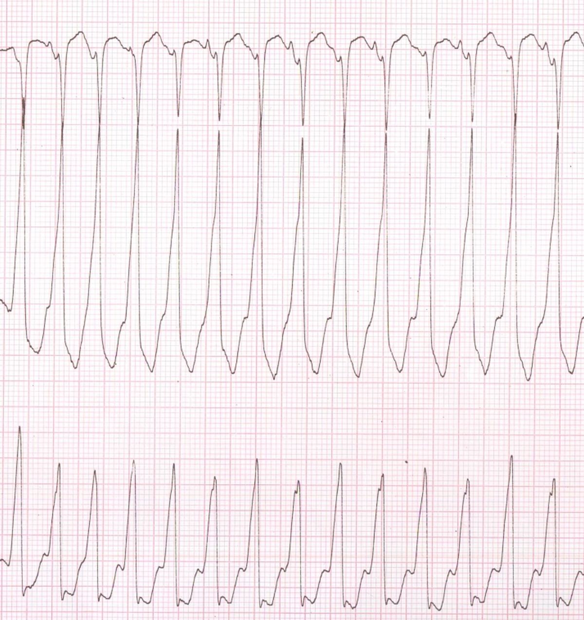
If the complexes have a uniform morphology in the same lead it is known as monomorphic VT. If the pattern varies in the same lead it is known as polymorphic VT. Monomorphic VT is often amenable to radiofrequency catheter ablation while polymorphic VT is not. VT occurring in the presence of structural heart disease generally has a poorer prognosis than those in individuals with structurally normal heart.
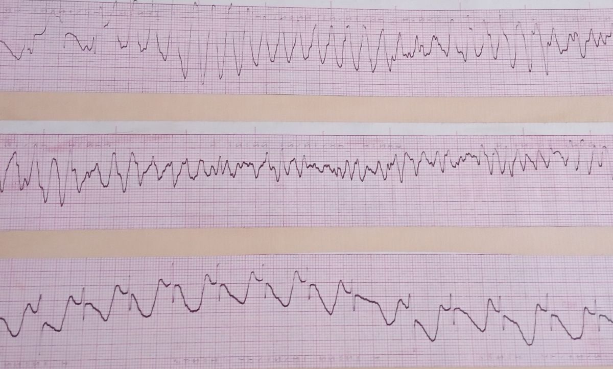
In the electrocardiogram, VT is characterised by wide QRS complexes at a rate more than 100 per minute. Some irregularity of rhythm may occur, especially in polymorphic VT. Irregularities can occur in monomorphic VT due to capture beats and fusion beats. Fusions beats have a morphology intermediate between that of the tachycardia beat and sinus beat. Dissociated P waves, if visible will pinpoint the diagnosis of VT in a wide QRS tachycardia. Presence of capture beats and fusion beats also enhance the diagnostic accuracy. Various morphological criteria including the Brugada criteria are useful in differentiating VT from other forms of wide QRS tachycardia.
Related Posts


About The Author
Johnson Francis
Former Professor of Cardiology, Calicut Govt. Medical Kozhikode, Kerala, India. Editor-in-Chief, BMH Medical Journal