PQRST in ECG
PQRST in ECG
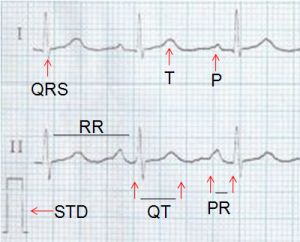
The waves and intervals in a normal ECG are illustrated here. P wave is due to atrial depolarisation. QRS is the ventricular depolarisation and T wave represents ventricular repolarisation. Atrial repolarisation is called Ta wave. Ta wave is not visible because it is shallow and superimposed on the PR segment, QRS and part of the ST segment. STD: standardisation pulse, a square wave of 1 mV amplitude, giving 10 mm vertical amplitude in usual ECG.
1 mm on X-axis represents 40 ms as the ECG is recorded at a speed of 25 mm/s. 1 mm on the Y-axis represents 0.1 mV. RR interval is the distance between the onset of two consecutive RR waves. 1500 divided by RR interval in mm gives the ventricular rate. PR interval is measured from the onset of P wave to the onset of the QRS and the normal range is between 120 to 200 ms in adults.
QT interval is measured from the onset of QRS to the end of T wave. It represents the systole of the ventricle and includes the depolarisation and repolarisation of the ventricle. PR segment is the near isoelectric region between the end of the P wave and the beginning of the QRS. ST segment is the region between the end of the QRS and beginning of the T wave. ST segment has a gradual upward slope in most ECGs. The true isoelectric interval in the ECG is the TP segment between two activations. But it need not be a straight line due to mild wandering of the baseline in most ECGs.
Related Posts


About The Author
Johnson Francis
Former Professor of Cardiology, Calicut Govt. Medical Kozhikode, Kerala, India. Editor-in-Chief, BMH Medical Journal