Patent Ductus Arteriosus (PDA)
Patent Ductus Arteriosus (PDA)
Patent ductus arteriosus is a communication between the pulmonary artery and aorta. Ductus arteriosus is a structure present in fetal life and its function is to shunt relatively deoxygenated blood coming from the upper part of the body to the descending aorta. This blood reaches the placenta for oxygenation. After birth, when the infant takes the first breath, pulmonary vascular resistance falls rapidly and pressures in the pulmonary artery falls. This leads to reversal of blood flow from aorta to pulmonary artery.
The mature ductus responds to high oxygen content of blood from the aorta after birth and starts constricting. Functional closure of ductus occurs within 72 hours of birth. Full anatomical closure takes a little longer. In premature infants, the ductus does not respond to oxygen and remains open. That is why premature babies have more incidence of PDA. The more the prematurity, the higher the chance of PDA.
Though the premature ductus does not respond to oxygen, it is responsive to prostaglandin. Prostaglandin keeps the ductus patent. Prostaglandin inhibitors like indomethacin, mefenamic acid and even paracetamol can be used to close the ductus pharmacologically in premature infants. Indomethacin is being less favoured now due to adverse effects. This does not work in mature infants.
In certain complex congenital heart diseases, it is essential to maintain the patency of the ductus to improve oxygenation. In such instances, patency of the ductus is maintained by giving a continuous infusion of prostaglandins. This is useful in both ductus dependent pulmonary circulation and ductus dependent systemic circulation. Sometimes PDA may be stented in such cases, to maintain patency.
PDA produces left to right shunting of blood and causes dilatation of the left atrium and left ventricle. If the shunt is large, it can cause heart failure in infancy. In the long term, shunt through the PDA leads to pulmonary hypertension and irreversible pulmonary vascular obstructive disease. When the pulmonary hypertension is severe, it causes reversal of shunt across the ductus (Eisenmenger Syndrome).
A patent ducutus arterious which is persisting beyond the immediate neonatal period has to be closed either surgically or by interventions. Triple ligation was a method of surgical closure while division and suture was another method.
But now-a-days the trend is towards interventional closure. Interventional closure can be done using coil embolization or device closure. Amplatzer device closure is a popular method, especially for larger PDAs. Intermediate sized PDAs can also be closed using simultaneous multiple coil delivery. Various types of coils with and without delivery systems were available.
Coils could also be delivered using a bioptome. This gave a better control for positioning the coil in the ductus before release. Rarely a coil would embolize either into the aorta or into the pulmonary artery. Embolized coils could be retrieved from major vessels using snares. Sometimes a coil which had embolized into a small branch of a pulmonary artery would also be left alone.
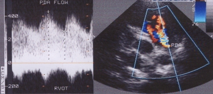
Left panel: Doppler pattern of PDA showing continuous flow – above the baseline, with a peak velocity of 400 cm /sec (left)
Right panel: Colour Doppler image showing multicoloured mosaic jet of PDA (right)
Related Posts


About The Author
Johnson Francis
Former Professor of Cardiology, Calicut Govt. Medical Kozhikode, Kerala, India. Editor-in-Chief, BMH Medical Journal